在消化疾病诊疗版图中,内镜既是“眼睛”,也是“双手”,更正在成为融合创新技术、精准诊疗与智能装备的重要平台。CGC2026会议期间,消化内镜板块将围绕领域的新技术、新理念与新边界展开,共探内镜创新与精准诊疗新方向。
胰腺疾病的超声内镜诊治进展
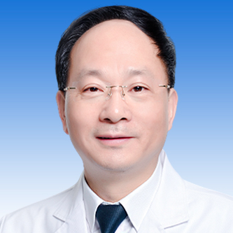
金震东
海军军医大学第一附属医院
近年来,超声内镜(EUS)在胰腺疾病诊疗中的应用不断拓展,已从单纯的影像学检查逐步发展为集精准诊断、标本获取与微创治疗于一体的综合诊疗手段,其应用范围涵盖EUS活检、EUS引导下消融、胰管引流及胰腺周围液体积聚治疗等多个方面。
在诊断层面,EUS引导下细针穿刺抽吸术和活检术(EUS-FNA/FNB)已成为胰腺实性病变获取病理标本的重要手段,尤其适用于影像学表现不典型、疑似自身免疫性胰腺炎或淋巴瘤、拟行新辅助治疗以及需要明确病理诊断的患者。经EUS-FNA/FNB获取的标本量可满足二代测序(NGS)检测需求,为胰腺恶性肿瘤的分子分型和精准用药提供依据。对于胰腺囊性病变,EUS能够清晰观察囊壁结节、分隔、囊内容物等细微结构,必要时可联合细针活检或囊液分析,进一步提高诊断准确性。组织学、细胞学、快速现场评估(ROSE)、肉眼现场评估(MOSE)及人工智能辅助评估等技术的发展,有助于提高取材质量和诊断效率。
治疗方面,EUS引导下消融术为部分患者提供了新的微创治疗选择。对于不适合手术或拒绝外科手术的胰腺囊性肿瘤患者,EUS引导下乙醇、聚桂醇及射频消融可作为外科治疗的重要补充;对于病灶体积较小的胰腺神经内分泌肿瘤,EUS引导下射频消融或乙醇注射具有创伤小、恢复快等优势;对于局部晚期或不可切除胰腺癌,也可探索EUS引导下射频消融,但应严格掌握适应证,并经多学科评估;对于胰管梗阻,EUS引导下胰管引流术(EUS-PD)可作为ERCP失败后的重要补救手段;对于胰腺假性囊肿(PPC)和包裹性坏死(WON),EUS有助于判断病变性质、坏死比例及囊壁结构,为支架置入和后续清创提供指导,EUS引导下的穿刺引流术是PPC和WON首选治疗方法。腔内贴合金属支架(LAMS)的应用,则进一步提升了引流效率和治疗安全性。
随着人工智能、分子病理和新型介入器械不断发展,EUS将在胰腺疾病的早期诊断、精准取材和微创治疗中发挥更加重要的作用。后续应在规范培训、精准取材、严格把握适应证及多学科协作的基础上,推动相关技术的合理应用。
主题:大消化·时代最强音 内镜-1
时间:9月18日 13:50-14:10
地点:102室
从柔性牵引到一体集成:消化内镜机器人辅助切除演进之路
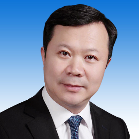
李锐
苏州大学附属第一医院
传统内镜下黏膜剥离术(ESD)面临黏膜下剥离视野受限、出血与穿孔风险高、学习曲线陡峭等结构性瓶颈。从调整体位借助重力辅助,到运用“黏膜桥”法、口袋法利用液体浮力,再到经皮牵引、磁控牵引、带线钛夹等各类新兴牵引手段,第一代牵引技术完成了从“顺应重力”到“创造引力”的主动革新,但本质上仍局限于单通道操作模式,操作三角缺失这一问题依然存在。
柔性机械臂的出现实现了机器人化牵引的关键跨越。以全球首个获批上市的消化内镜手术机器人EndoFaster为例,其微型连续体柔性机械臂直径约3 mm,可在消化道有限空间内实现4自由度精准主从控制。多中心随机对照试验结果显示,EndoFaster辅助ESD可显著短于总手术时间(18.8 min vs 32.8 min),直视剥离率由73%提升至96%,肌层损伤明显减少。针对学习曲线的研究进一步提示,该技术能缩短新手学习周期、降低操作门槛。李锐教授团队已成功应用该技术,先后完成食管静脉曲张背景下食管黏膜病变的剥离、胃体前壁双病灶的直视下剥离、十二指肠巨大侧向发育型肿瘤的完整剥离及困难直肠病变的完整切除;同时牵头成立了国内首家消化内镜手术机器人培训示范基地。
放眼一体集成式内镜机器人的发展演进图景,多臂主从协同系统有望重塑内镜下的操作三角,推动手术范式从“一手扶镜、一手切割”转向双手协同操作。同时,结合力触觉实时反馈、术中人工智能(AI)辅助决策与自主导航、多模态影像融合成像等功能,系统正逐步构建起“感知-决策-操作”的完整闭环。“一体集成”的终点并非硬件集成的完成,而是智能化一体平台的收敛——硬件提供操作平台,AI提供决策能力,多模态感知提供精准眼界。
随着机器人辅助技术走向规范化、标准化,内镜切除的适应证有望进一步拓展,学习曲线缩短,内镜手术流程化、标准化与“技术平权”的时代正在到来。
主题:大消化·时代最强音 内镜-3
时间:9月18日 15:50-16:05
地点:102室
Innovation and Emerging Technologies in Digestive Endoscopy
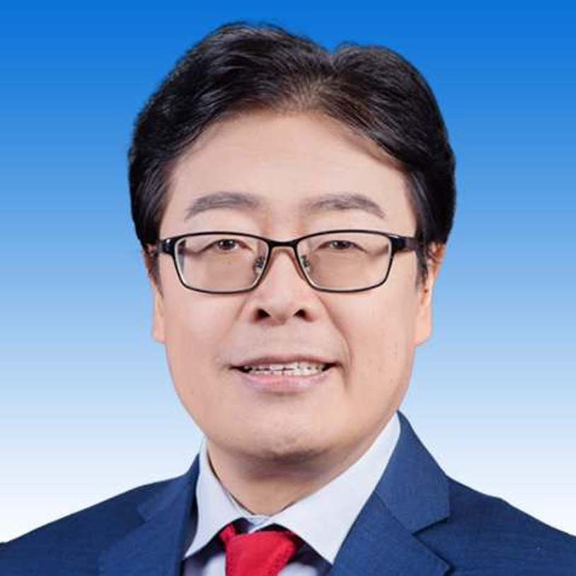
李鹏
首都医科大学
附属北京友谊医院
In 1805, Bozzini in Germany used candlelight through a rigid tube to inspect the rectum. That was the first endoscope: crude, rigid, dangerous.
In 1868, Kussmaul built the first straight-tube gastroscope. In 1932, Schindler's semi-flexible gastroscope appeared; the shaft could bend, and the working range opened up at once.
Two centuries, two themes: greater flexibility, greater illumination. Then electronics replaced optics.
Early GI cancer
Early cancer is where endoscopy proves its value most consistently.
The key word is quality. Screening only matters if it is done well. Miss an advanced adenoma, and five years later you may be facing an interval cancer.
Whole-chain management
Our work in early cancer is not simply resection. President Zhang Shutian proposed the concept of whole-chain management. Whole-chain management, multidisciplinary collaboration, and systematic coordination are the only viable path to delivering early GI cancer diagnosis and treatment.
The ideal is that no one develops cancer at all, which is why we do public education. If they do develop it, they must come early. Early cancer is curable.
The chain does not end at resection. It continues through standardized specimen handling, pathology reporting, and follow-up. That is what whole-chain means.
Improving ADR
A concrete quality metric for colonoscopy screening: the adenoma detection rate, ADR.
There are two ways to improve it. One is to see the mucosa better, with image-enhanced endoscopy: NBI and TXI on the device side, indigo carmine and crystal violet on the dye side. The other is to widen the field of view, with 3D endoscopy such as FLEX 3D and auxiliary devices: transparent caps, Endorings, Endocuff, AmplifEYE, WingCap, Embrella. The principle is the same in every case: unfold the folds.
From white light to image enhancement
After white-light endoscopy came image-enhanced endoscopy: NBI, BLI, TXI. Diagnosis has extended into therapy; a single platform both detects and removes lesions. The RDI function helps identify vessels, making resection more precise.
AI for quality control
Now, AI. Its use in detecting colonic polyps and adenomas is already increasingly widespread.
This is a multicenter randomized controlled trial published in *Endoscopy*. The team built an AI system that monitors quality during esophagogastroduodenoscopy.
The study moves AI into quality control of upper GI endoscopy. AI is now fully integrated into digestive endoscopy screening.
See clear, see accurately, see precisely
Modern endoscopic imaging in three phrases: see clear, see accurately, see more precisely.
See clear means high resolution and 4K. See accurately means image enhancement and magnifying observation. See precisely is confocal laser endomicroscopy, which gives us an optical biopsy: cellular-level information, in vivo and in real time.
AI-assisted ESD
AI is not only for examination. In ESD it now assists with three tasks: marking, vessel identification, and dissection plane guidance. The work was published in the *International Journal of Computer Assisted Radiology and Surgery*, and more recently by Scheppach and colleagues in *Endoscopy*.
On the operating table: marking defines the boundary, vessel identification indicates where bleeding risk lies, and dissection plane guidance keeps us in the correct layer.
These three tasks are exactly where young endoscopists struggle most. AI shortens the learning curve and makes treatment more precise.
Sentinel lymph node navigation surgery
Treatment of early GI cancer is also innovating. How do we save more gastric function in the grey zone between ESD and standard surgical treatment for early gastric cancer?
Sentinel lymph node navigation surgery for early gastric cancer. The core issue is balance: oncologic safety against organ function preservation.
The logic is straightforward. If the sentinel node is negative, the other nodal regions are considered negative as well. We can therefore avoid extensive lymphadenectomy, preserve function, and at most perform local gastrectomy.
A meta-analysis of 38 studies reported a sentinel node detection rate of 94% and accuracy of metastasis prediction of 92%.
The procedure: indocyanine green injection, lesion marking, incision around the margin, dissection, completion of ESD, and observation of the tracer after 15 minutes.
T1 colorectal cancer
For T1 colorectal cancer, the question concerns expanding endoscopic indications. Where is the boundary of endoscopy? For lesions suspected of T1b invasion, we must ensure negative margins where possible, perform full-thickness resection when needed, and add chemoradiotherapy when needed. For elderly patients, this is an important option for sphincter preservation and avoiding surgery.
Advanced rectal cancer
A frontier worth watching: chemoradiotherapy combined with ESD for advanced rectal cancer.
The driver is sphincter preservation. In this patient with advanced rectal cancer, downstaging after chemoradiotherapy was marked, and ESD was then performed.
Endoscopy is gradually extending its reach. The boundary with surgery is becoming increasingly blurred.
Innovation in closure
Now, closure. Resection is only half the story; the other half is closing the wound safely.
Eight-ring suturing and double-layer suturing, from *Therapeutic Advances in Gastroenterology* in 2025. This slide shows a three-arm grasper closing a large wound, from *Surgical Endoscopy* in 2024. Further along, innovation in the suturing systems themselves: handheld suturing systems, Overstitch, and others.
The direction is consistent: from simple clips toward full-thickness, secure, adjustable suturing.
ERCP and EUS
Now the pancreaticobiliary system, and the integration of ERCP and EUS.
In the past we could only address stones within the common bile duct. Today the indications are expanding: ERCP-guided transpapillary gallbladder drainage has grown into transcystic gallbladder stone extraction.
This is the development of gallbladder stone extraction. Early on it was bridging drainage for patients with high-grade acute cholecystitis, demonstrating that we could reach the gallbladder through the papilla. The technique then accumulated: anatomical classification of the cystic duct, with cannulation success rising to 85–90%. In 2021 the four-step ETGBD classification was published, and SpyGlass-assisted success rose from 72% to 94%. In 2023, a complete video workflow for cholangioscopy-guided transcystic gallbladder-preserving stone extraction was published, making the technique reproducible. Today, indications have moved from drainage toward therapy.
Five steps: selective biliary cannulation; cholangioscopic identification of the cystic duct orifice with guidewire placement; advancement into the gallbladder over a bridge stent; gallbladder irrigation; and finally stone extraction or lithotripsy.
Interventional EUS
The other component is interventional EUS, which began with EUS drainage of pancreatic pseudocysts. That was the dividing line where EUS stopped being purely diagnostic.
We now call it interventional EUS. We propose five categories:
One, EUS-guided sampling. Two, EUS-TTNE, trans-needle examination. Three, EUS-D/A, drainage and anastomosis. Four, ESCR, EUS-created-route surgery. Five, EUS-guided delivery.
Take EUS-TTNE, trans-needle examination. Three subcategories. Imaging: EUS-guided confocal laser endomicroscopy and EUS-guided cystoscopy. Measurement: portal pressure gradient, EUS-PPG, and impedance spectroscopy. Biopsy: EUS-TTNB, sampling through the needle tract with micro forceps such as the Moray device.
This is a new space, and it will develop rapidly.
EUS-BD, EDGE, and the liver
Clinically, the most important advances are in drainage and access.
EDGE, EUS-guided gastrostomy-assisted ERCP, is for patients with surgically altered anatomy. Under EUS guidance we create a gastrostomy, inject contrast or carbon dioxide into the remnant stomach, place a self-expanding metal stent to dilate the tract, and then pass a duodenoscope to complete standard ERCP. The stent is removed at the end.
Then EUS-BD, the option after failed ERCP, which divides into several routes: EUS-CDS, choledochoduodenostomy; EUS-HGS, hepaticogastrostomy; EUS-RV, the rendezvous technique; EUS-AG, antegrade treatment; and EUS-TASR, antegrade stone removal.
EUS is also entering hepatology: liver biopsy, elastography, intra-variceal coil and glue injection, portal pressure measurement, and portal vein sampling.
EUS-GE
Similarly, consider the development of EUS-guided gastroenterostomy, EUS-GE.
The concept of endoscopic gastrojejunostomy was first proposed in 1991, and the EUS-GE concept followed in 2002. In 2012, LAMS, lumen-apposing metal stents, were first used. In 2013 the first EPASS procedure was completed. The field has expanded since.
For malignant gastric outlet obstruction, EUS-GE is now beginning to compete with surgical bypass.
Third-space endoscopy
Next, functional disorders, and third-space endoscopy for benign motility disorders.
Four procedures share the same logic: submucosal injection, tunnel creation, myotomy, and clip closure.
First, POEM+E, POEM combined with fundoplication, which addresses POEM's greatest weakness: post-procedure reflux.
Second, G-POEM, for gastroparesis. The tunnel technique applied to the pylorus.
Third, Z-POEM, for Zenker's diverticulum, with selective myotomy of the cricopharyngeal muscle.
Fourth, per-rectal endoscopic myotomy, PREM, for Hirschsprung's disease.
GERD and the MUSE system
For GERD we have several options: radiofrequency therapy, LinX, ARMS, and laparoscopic fundoplication.
MUSE is an intraluminal surgical platform that operates through natural orifices. Miniature imaging, ultrasonic detection, surgical anastomosis, and intraoperative localization are integrated within it. It is used to treat GERD.
The endoscope is inserted transorally, advanced through the esophagus, and retroflexed in the gastric fundus. The fundus is stapled to the lower esophagus at two or more points, creating a partial fundoplication. Specifically, stapling is performed along the greater curvature 3 cm above the cardia. By creating a 150–180 degree anterior partial fold, the procedure raises cardial pressure, restores the angle of His, and reconstructs the gastroesophageal junction.
No incision, completed through the mouth. The domestic VES-800P stapler and CV-H800I host system make this possible.
Bariatric endoscopy
Next, bariatric endoscopy.
The mechanisms fall into three groups: restricting gastric volume and delaying emptying, which includes intragastric balloons, suture plication such as endoscopic sleeve gastroplasty, and electrical stimulation; promoting clearance of gastric contents, that is, gastric aspiration therapy; and reducing intestinal absorption, that is, duodenal-jejunal bypass and duodenal mucosal resurfacing.
Three mainstream approaches currently: the intragastric balloon, endoscopic sleeve gastroplasty, and the gastric bypass stent.
ESG was proposed in 2008 and first performed in 2012. It is currently the most mature endoscopic bariatric procedure. The mechanism is volume restriction with delayed emptying. Based on the Overstitch system, continuous and interrupted full-thickness sutures are placed along the greater curvature from within the lumen. Gastric volume decreases, satiety comes earlier, and gastric emptying and hormone secretion change accordingly. The name derives from its resemblance to surgical sleeve gastrectomy.
We now have a single-use suturing system, the first suturing device in China approved for ESG. Continuous full-thickness suturing draws tissue together and tightens it. There is no incision, and gastric anatomy remains intact. It fits the vast majority of single-channel endoscopes, can close large GI defects, and the suture path can be set as needed.
Endoscopy robots
Finally, robotics.
This is a single-arm flexible robotic system for natural orifice surgery. It can be assembled onto endoscopes of any brand, making installation convenient. The platform supports multiple consumables, so one system covers different procedures.
Compare the ESD robots currently in development. China's EndoFaster is a traction-assistance system for gastric and colorectal ESD. China's EndoDreams is a GI intraluminal surgical platform. Singapore's EndoMaster is an intraluminal dual-arm surgical robot. America's EndoQuest targets rectal and sigmoid lesions. Korea's EndoRobotics is an endoscopy-assist system.
They differ in dual-arm coordination, endoscope control, and compatibility with routine workflows, each targeting a different clinical niche.
Robotic-assisted ERCP has also entered exploratory studies.
Capsule endoscopy
Capsule endoscopy is moving toward capsule robotics. This image shows a beef tapeworm, captured by capsule in the small bowel.
Our innovative capsule endoscopy system can achieve three-dimensional panoramic reconstruction: no longer a stack of planar images, but a complete spatial model of the entire intestinal lumen. This has significance for automated lesion localization and AI reporting. The video demonstrates dynamic images from an intraluminal perspective.
Conclusion
The diagnosis and treatment of early GI cancer is moving toward whole-chain, multidisciplinary management. AI is fully integrated and participates in quality control.
From white light to NBI and TXI, and on to confocal endomicroscopy. Imaging is moving toward the microscopic, becoming ever more refined.
Resection and closure continue to advance: AI-assisted ESD, and new full-thickness suturing systems.
ERCP and EUS have merged into a shared interventional platform, reaching organs we could previously only image.
Third-space endoscopy has extended the tunnel from the esophagus to the rectum, offering solutions for motility disorders.
Bariatric endoscopy is becoming a first-line option.
Digestive endoscopy robots and capsule robots point toward the future.
Underlying all of it is a persistent search for solutions: to accomplish through natural orifices, with minimal injury, what once required open surgery.
主题:消化领域新锐领导者联合-趋势与研究挑战
时间:9月17日10:20-10:40
地点:101室
(以上讲题、时间及地点安排以大会最终实际日程为准,如有调整,恕不另行通知。)
欢迎投稿!
tougao@medvoices.cn
稿件类型:研究速递,综述译稿,指南/共识,病例分享。